Peripheral-Artery-Disease

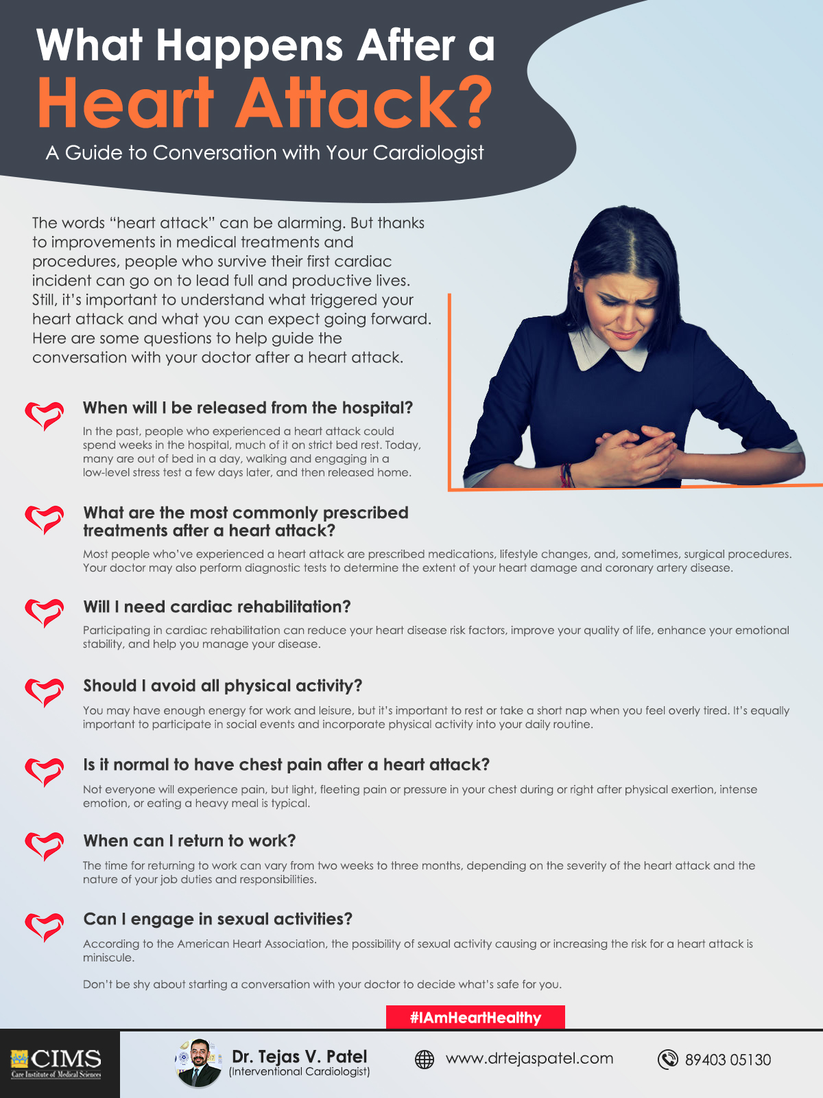
Leg Pain While You Walk? Never Ignore It

Walking is considered as one of the best exercise for a better health. But what if your leg causes pain, which is not a part of your aging?

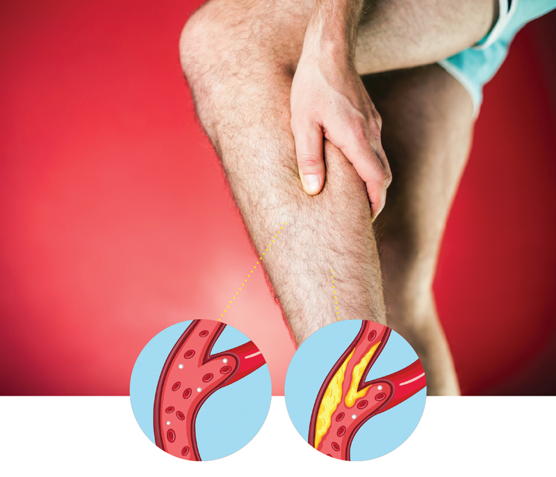
Many people ignore the leg pain as a normal sign of aging. Sometimes you may think it’s arthritis, sciatica or just “stiffness” from getting older. In some cases, it is the sign of Peripheral Artery Disease (PAD), putting your heart and brain health at greater risk.

While PAD doesn’t usually run in families, it occurs due to various reasons like aging, smoking, having high blood pressure, high cholesterol or diabetes. If you have any of the risk factors for PAD, you should ask your doctor about PAD even if you aren’t having symptoms yet.

What causes leg pain when you have PAD?

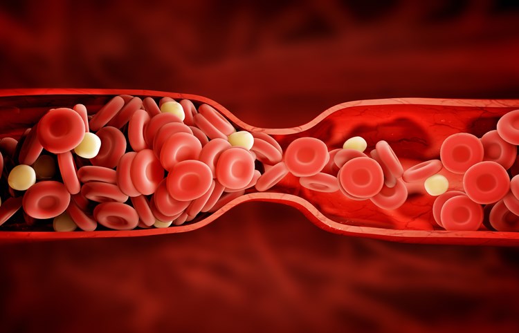
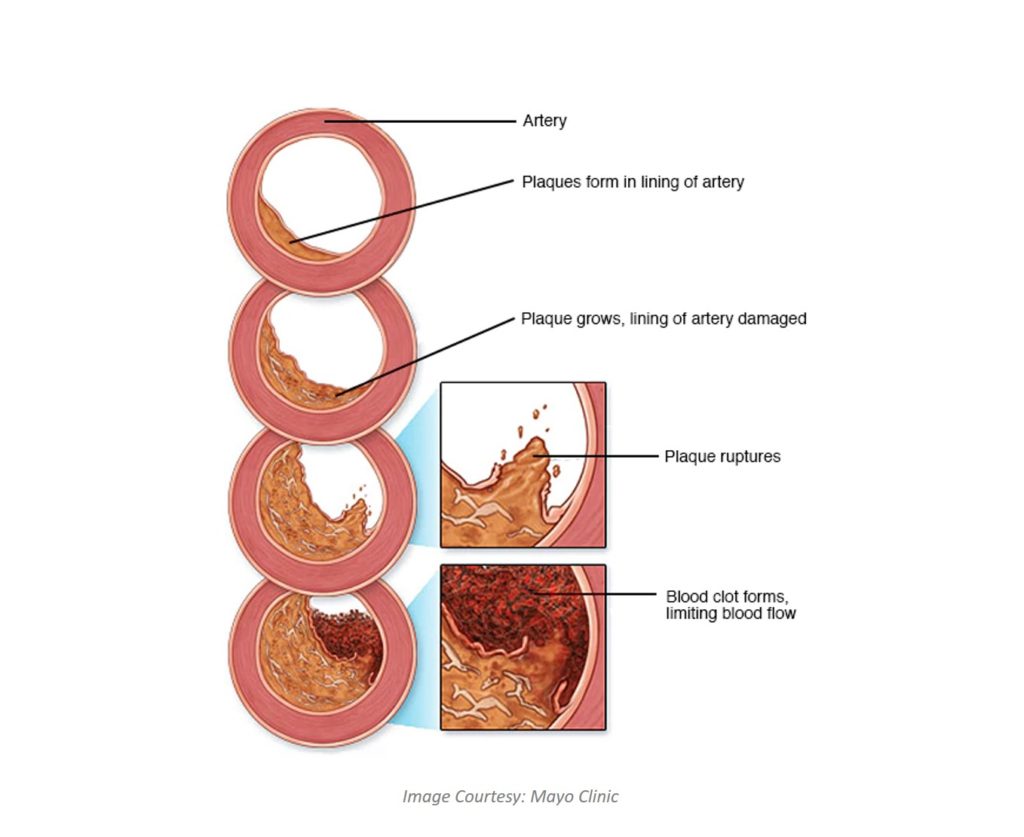
People with PAD have fatty deposits in the arteries outside the heart, usually in their legs. When these deposits block blood flow to the muscles, impairing their ability to work properly, it causes pain.

Initially it was thought that PAD occurs mostly in men, but later study found that the condition is just as common in women affecting one in every 10 women over age 50. [Source: Harvard Health Publishing]

Symptoms of Peripheral Artery Disease (PAD)

Leg pain is a very common symptom of having PAD, but not everyone has the same symptoms. Some may experience just weakness without any pain or cramping in leg. Yes, it has the same pattern that is worsening with exercise or movements and easing with the rest.

In some cases, people notice other changes such as:

  • Slow healing sores on the feet
  • Coldness in one of both feet
  • Slow or poor growth of toenails or leg hair
  • Gangrene, or dead tissue
  • Leg pain that does not go away when you stop exercising

Why you can’t ignore the leg pain while walking

Of course PAD is not the only reason of leg pain, but it’s important to consider at the same time. Having PAD increases your risk of other cardiovascular diseases. This makes a person with PAD far more likely to have a heart attack or a stroke than someone without the condition.

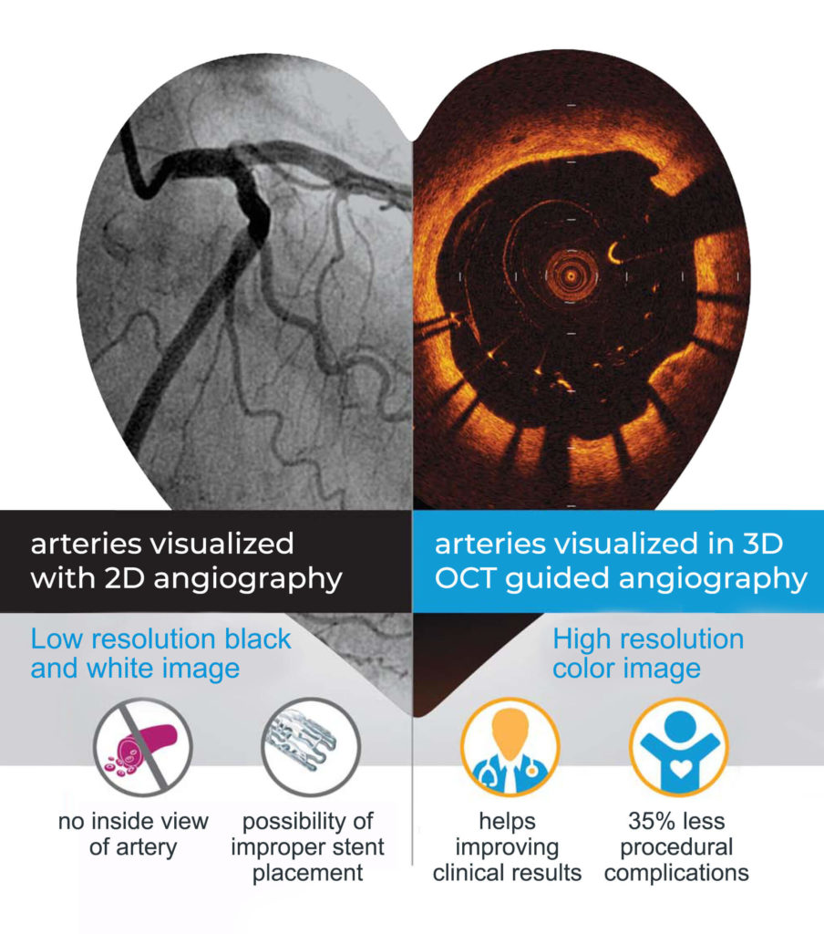
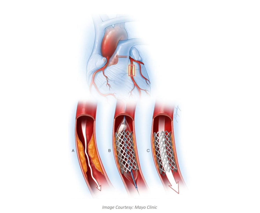
If your doctor suspects you have PAD, you might want to follow up with a peripheral angiography, which uses MRI or x-rays to take images of your arteries to look for blockages.

Treating PAD with lifestyle changes and more…

PAD treatment always starts with a healthy lifestyle that includes avoid smoking and other tobacco products, regular exercise, healthy diet rich in fruits and vegetables and consuming healthy fats.

One of the most important is ignoring the leg pain thinking it as a part of getting old. That’s exactly what you should not do. Talk to your doctor immediately and find out why it occurring.

In addition to lifestyle changes, your doctor may also prescribe some medications to treat PAD. Most common drugs prescribed for people with PAD are to keep more fatty deposits (plaque) from accumulating, preventing blood clots and controlling high blood pressure.

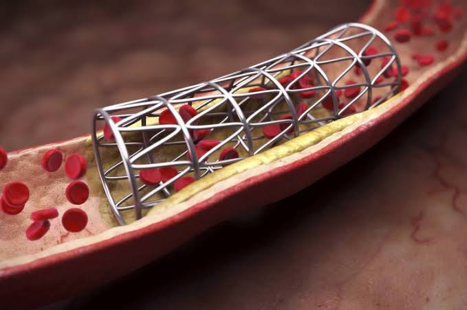
If you are diagnosed with severe blockage, your doctor may also recommend a procedure to clear the blockage or to reroute blood flow around it. Always remember, “the longer you wait, the harder it is to treat.

Categories:
oct_main

OCT & FFR

It is a new, non-invasive imaging technique that generates volumetric angiography images in a matter of seconds.

OCT & FFR

OPTICAL COHERENCE TOMOGRAPHY (OCT) GUIDED ANGIOPLASTY

Optical coherence tomography (OCT) is a diagnostic procedure that is used during cardiac catheterization. It is a new, non-invasive imaging technique that generates volumetric angiography images in a matter of seconds.

OCT uses light and with OCT, doctors can obtain images of the blood vessels that are about the same as if they were looking under a microscope.

HOW DOES OCT WORK?

OCT uses near-infrared light to create images of the inside of the coronary arteries. The technique delivers very high-resolution images. In fact, OCT allows cardiologists to see the inside of an artery in 10 times more detail with 3D image than traditional angiography with 2D images.

BENEFITS WITH OCT

oct_benefits_tiny
OCT also lets cardiologists clearly see the plaque inside an artery, find out how much fat or clot is inside an artery, and take precise measurements before and after placing stents.

FRACTIONAL FLOW RESERVE (FFR)

Fractional Flow Reserve (FFR) is a procedure to determine if a cardiac patient really needs a stent or bypass surgery or can be kept only on medicines avoiding any procedure. FFR technology not only saves lives while avoiding unnecessary surgery but also helps patients to save cost.

HOW DOES FFR WORK?

A very thin guide wire is inserted through a standard 4F or 5F diagnostic catheter during an angiogram. Because of the smaller size catheter necessary, this can be done as an outpatient procedure.

The special guide wire crosses the lesion and is able to measure the flow and pressure of the blood, after infusion of a hyperemic agent, such as adenosine. Results are displayed on a special monitor (left) along with the “FFR value”. [Source: PTCA.ORG]

Need more information about OCT & FFR, talk to us now.

Blood-Clots

Symptoms of Blood Clotting – How to Tell If You Have One

Ever got a knife cut on your finger or a blade’s cut while shaving? When that happens, a blood clot saves the day as it quickly stops bleeding. Proteins and particles in your blood, called platelets, stick together to form the blood clot. after it has done its job, it breaks up.

Though sometimes things can go wrong. The blood shouldn’t clot when it’s just moving through the body. If blood tends to clot too much, it is referred to as a hypercoagulable state or thrombophilia. They can be dangerous and lead to serious medical conditions.

They are most likely to affect a leg, when you sit for long durations.
You also get a clot in your arteries, which carries oxygen in your blood from your heart to all the cells of your body. The result can be really serious and cause a life-threatening emergency, like a heart attack or stroke.
You could also get a clot in the veins that carry blood back to your heart.

We have listed out a few warning signs. Scroll down to learn more and you’ll know when to get a medical help.

ARMS & LEGS

Deep Vein Thrombosis (DVT) – is when a blood clot forms in one of the deep veins in your arm or leg. This is dangerous and this clot could travel to your heart. Get medical help right away if you notice any of these symptoms:

  • Arm or leg swelling
  • Change in colour – red or blue tinge
  • Dull ache to intense pain
  • Trouble breathing
  • Lower leg cramp

HEART

A blood clot around your heart may cause a heart attack. Watch out for these symptoms:

  • Sweating
  • Severe pain in your chest
  • Trouble breathing

LUNGS

A blood clot in your lungs usually starts with a clotting in your arm or leg, which travels to your lung. This is extremely dangerous condition, so get medical assistance if you:

  • Feel short of breath
  • Pain in your chest
  • Cough
  • Sweating
  • Dizziness

BRAIN

Blood clots here may be caused by fatty deposits in the walls of the blood vessels that bring blood to your brain. In other cases, it can also be occurred when a clot that starts out in a different part of your body, like your chest or neck, enters your bloodstream and move to your brain, where it can cause a stroke.

Watch out for these symptoms:

  • Seizure
  • Feeling weakness
  • Vision problem
  • Speech problem

OTHER AREAS

Belly – Blood clots can happen in the veins that drain blood from your intestines. They can be caused by conditions like diverticulitis or liver disease, or even by birth control pills.

Kidneys – A blood clot there can keep them from removing waste from your body. That can cause high blood pressure or even kidney failure.

It’s important to call or contact cardiologist right away if you have any symptoms of a blood clot. Getting prompt treatment will help prevent any potential complications.

Categories:
images

Healthy Lifestyle Can Ease the Risk of Genetic Heart Disease

Is heart disease or stroke in your family? If so, your risk may be higher.

Cardiovascular disease is the #1 cause of death worldwide. So most of us may have a family history of, and some genetic risk for, heart disease.

Did your father have a stroke?
Did your mother have a heart attack?
Did any of your grandparents have any heart disease?

Yes, these are the random questions your cardiologist may ask, and they are very important when it comes to understanding your risk factors for heart diseases. Because knowing your family history can help you avoid heart disease.

So I’ve got family history concerns … what next?


Just because your family has a history of heart diseases, doesn’t mean you will certainly have the same disease. It just means you are more likely to have them.

Diseases are not imminent and your health can be managed by making healthy lifestyle choices. Healthy habits can cut your risk of heart disease in half, even if your genes are stacked against you, according to new research.

“People with the highest genetic risk scores had nearly twice the risk of having a heart attack or a related problem as those with low scores. But people who had three or four of the healthy lifestyle habits cut their risk in half, compared with people who had none or just one of the habits, the researchers found.” [Source: Harvard Health Publishing]

Simplest Healthy Lifestyle Options


Healthy Diet – including more fruits and vegetables, whole grains, nuts, fish, limited intake of sodium, added sugars and saturated fats.

Regular Exercise – at least once per week. Even a long brisk walking will do the job.

No Smoking – avoid any type of smoking habits, not even the electronic cigarettes. [Know what happens when you quit smoking.]

No Obesity – as defined by body mass index (BMI)

Control your numbers – both high blood pressure and high cholesterol levels increase the risk of a heart attack. You can keep them in check with lifestyle changes and, in some cases, medication.

Ending Note…


We know that both genetic factors/family history and lifestyle choices contribute to the development of heart disease. Further your risk of heart disease can be lowered through a healthy lifestyle changes that only you have the power to change.

Skip blaming your mom or dad (they didn’t ask for this family history, either) and take action instead.
Choose a healthy lifestyle and say #IAmHeartHealthy.

Categories:
angioplasty_main

Coronary Angioplasty & Stents

OVERVIEW

Coronary angioplasty, also known as percutaneous coronary intervention, is a procedure to open clogged heart arteries. This procedure involves inserting and inflating a tiny balloon, exactly at the place where your artery is clogged, to widen the artery.

Angioplasty is often combined with the permanent placement of stent (a small wire mesh tube), to prop the artery open and decrease its chance of narrowing again. This procedure or treatment can improve symptoms of blocked arteries, such as chest pain and shortness of breath. This helps quickly open a blocked artery and reduce the amount of damage to your heart.

WHY IS IT DONE?

Angioplasty treats atherosclerosis (slow build-up of fatty plaques in your heart’s blood vessels). Your cardiologist may suggest angioplasty as treatment when medications or lifestyle changes are no more enough to improve your heart health.
plaque
Again angioplasty is not for everyone. If the main artery is narrow or heart muscle is weak, then coronary artery bypass surgery may be a better option than angioplasty. Also if you are diabetic or have multiple blockages, angioplasty is not a treatment option for you.

WHAT YOU CAN EXPECT DURING ANGIOPLASTY

Angioplasty is performed by a cardiologist (heart specialist) and a team of specialized cardiovascular nurses and technicians in a special operating room called a cardiac catheterization laboratory. This room is often called the cath lab.

A very small incision is made on the skin over a blood vessel in the leg, arm or wrist. Catheter (thin tube) is threaded through the incision and the procedure is performed. It may take up to several hours based on number of blockages and any other complications.

Very commonly angioplasty is performed through an artery in your groin. Before the procedure the area is prepared with an antiseptic solution and a sterile sheet is placed over your body. Small electrode pads are placed on your chest to monitor your heart during the procedure. You’ll be sedated but awake during the entire procedure. You’ll receive fluids, medications to relax you and blood-thinning medications through an IV catheter.

STENT PLACEMENT

stent_placement
Most people who have angioplasty, will also have a stent placed in their blocked artery. The stent placed inside the artery supports the walls of artery and prevent from narrowing again.

After your stent placement, you may need prolonged treatment with medications, to reduce the chance of blood clots forming on the stent.

RESULTS – AFTER ANGIOPLASTY

Angioplasty with stent increases the blood flow through previously blocked artery. Your chest pain decreases gradually. Having angioplasty and stenting doesn’t mean your heart disease goes away permanently. You will have to strictly follow a healthy lifestyle and take proper medications as prescribed by your cardiologist.

A healthy lifestyle continuity will help you live a life normal and very soon you may be able to return to your routine. There are certain things that needs to be followed very strictly after angioplasty, such as:

  • Quit smoking
  • Lower your cholesterol levels
  • Maintain a healthy weight
  • Control other conditions, such as diabetes and high blood pressure
  • Get regular exercise

A successful angioplasty means you might not have to undergo a coronary artery bypass surgery. If any time your symptoms returns with chest pain or shortness of breath or any other symptoms you had earlier, contact your cardiologist immediately.

Categories:

Added Sugars – Cutting Back On All The Extra Sugar Is Not A Bad Idea

Sugar is a number one public enemy in nutrition and diseases these days. We don’t actually need an extra sugar than what is found naturally in whole foods. So cutting back on all added sugar foods is not at all a bad idea.

Believe it or not, if you can’t take out all the added sugars, just by reducing the intake, you will see the benefits. You will also struggle a little, as sugar can truly mess with your brain and act like an addictive substance.

What is a healthy diet and how much should I eat? This is the dilemma of every person on diet. Eating healthy can be easy, tasty and inexpensive if you follow some basic guidelines.

Find out more about what it means to cut out added sugar, where do the come from and how can you avoid them and ways you or your client can make this diet change healthfully and with minimal suffering. Scroll to see the infographic.

I256

Categories:

Dr Tejas V Patel

Area of Specialization

  • Radial angiography and angioplasty (& stenting)
  • Complex Coronary Interventions like- Rotational Atherectomy, Left Main coronary intervention
  • FFR Estimation, Coronary imaging – IVUS, OCT
  • Transcatheter aortic valve replacement (TAVR / TAVI)
  • Device closure of ASD, VSD, PDA
  • Other structural cardiac interventions- BMV, BPV
  • Peripheral Angioplasty- Renal Angioplasty, Carotid Angioplasty, Limb Angioplasty
  • Aortic aneurysm / dissection endovascular repair – TEVAR, EVAR
  • Permanent Pacemaker Implantation & AICD Implantation (Automated Implantable Cardioverter Defibrillator)

PROFESSIONAL EXPERIENCE

  • Consultant Interventional Cardiologist, Care Institute of Medical Sciences (CIMS) Hospital, Ahmedabad – June 2015 till date
  • Assistant Professor in Dept. of Cardiology, Christian Medical College (CMC) & Hospital, Vellore – Sept 2014 to May 2015
  • Senior Registrar as DM Trainee in Dept. of Cardiology, Christian Medical College (CMC) & Hospital, Vellore – August 2011 to August 2014
  • Registrar in Dept. of Medicine, M P Shah Medical College and Guru Gobind Singh Hospital, Jamnagar – 2006 to 2009

AWARDS & ACHIEVEMENTS

  • Having experience and part of Heart transplantation team at CIMS hospital (only hospital in Gujarat achieved 5 heart transplantation so far) – Did observership at UMPC Presbyterian hospital, Pittsburgh, USA (2016) & Global hospital, Chennai (2015)
  • Having experience of TAVR cases at CIMS hospital (only hospital in Gujarat has done successful TAVR cases) – certified by Medtronic Academy TAVR program
  • Have been Faculty & Course Director of Basic & Advanced Echocardiography Course by GE Healthcare Institute (2016 & 2017)
  • Have done CT coronary angiography course certified by Society of Cardiovascular Computed Tomography [2016, Fortis Escorts Heart Institute, New Delhi]
  • Awarded Associated Membership of Asia Pacific Vascular Society (2015)
  • Having experience for all complex peripheral interventions like TEVAR & EVAR (including fenestrated graft interventions) – trained under Dr George Joseph, CMC, Vellore
  • Was part of STEMI INDIA program [www.stemiindia.com/]at CMC, Vellore – was actively involved in development of similar program at CIMS hospital
  • Stood 1st in D.M. (Cardiology) exam held at CMC (2014) -awarded Dr G M Cherian gold medal for "Best Outgoing Student in DM Cardiology"
  • Selected for zonal round (among top 10) in Torrent Young Scholar Award (TYSA) competition during both M.D. [Mumbai, 2009] and D.M. [Bangalore, 2013]
  • Stood University 2nd in M.D. (General Medicine) exam held at Saurashtra University (May 2009)

EARLY LIFE AND BACKGROUND

Born in the historical land of Saurashtra, Dr Patel finished his school education from Junagadh. He completed his MBBS and MD (Medicine) from MP Shah Medical College, Jamnagar. He achieved 2nd rank in the Saurashtra University in MD.

Dr Tejas V Patel pursued his super-speciality, DM in Cardiology from Christian Medical College (CMC), Vellore; which is considered as one of the best medical institutions in India. He was awarded the prestigious “Dr G M Cherian Gold Medal” for ‘Best Outgoing Student in DM Cardiology’. He worked as Assistant Professor in this premier institute, after he finished DM. He spent almost four years at CMC, where he gained extensive experience in clinical, interventional and research work in the field of Cardiology.

Dr. Tejas V. Patel has wide experience of performing coronary angiographies and coronary angioplasties (majority performed through transradial approach). He is also trained for various peripheral vascular interventions including intervention in Takayasu arterities, Aortic aneurysm and dissection (EVAR and TEVAR procedures). Dr. Patel has independently performed various diagnostic and interventional cardiac procedures such as percutaneous ASD/VSD/PDA device closure, Balloon Mitral/Pulmonary/Aortic Valvulotomy and cardiac catheterization study with or without oxymetry of various congenital heart diseases.

He has done more than 10,000 transthoracic echocardiography including paediatric echocardiography, Tissue strain/strain rate echo, 3D echo and trans-oesophageal echo (TEE). As per the patients treated by him. He has been rated as one of the best cardiologist in Ahmedabad, Gujarat.


Dr. Tejas V. Patel has participated in several original research projects and many of his papers have been published at National and International levels in reputed journals including ‘Journal of American College of Cardiology (JACC)’. He has also given presentation at various national conferences. He was selected for zonal round (among top 10) in Torrent Young Scholar Award (TYSA) competition during both M.D. [Mumbai, 2009] and D.M. [Bangalore, 2013].

EDUCATION

  • Training at Seoul, Korea and Hong Kong University for advance technique in Angioplasty and stenting like OCT & FFR
  • Observership in advance Heart Failure Management and Heart Transplantation at University of Pittsburgh Medical Center (UPMC), Pittsburgh, USA
  • DM Cardiology: Christian Medical College, Vellore, Tamilnadu, India (between 2011-2014)
  • MD (Medicine): M P Shah Medical College & Guru Gobind Singh Hospital, Jamnagar, Gujarat (between 2006-2009)
  • MBBS: M P Shah Medical College, Jamnagar, Gujarat (between 1998-2004)
  • Secondary and Higher Secondary Education (School): 1996 passed 10th from Carmel Convent School, Junagadh, Gujarat & 1998 passed 12th from Swami Vivekanand High school, Junagadh, Gujarat

Research –Publications & Presentations

  • Invited as Faculty in India Live 2018 conference at Chennai for presentation in section ‘Meet the Expert – CD presentation’ ; presented- “Simple calcific RCA lesion – Never underestimate”
  • Invited as Faculty in India Live 2018 conference at Chennai for presentation in section ‘Meet the Expert – CD presentation’ ; presented- “Simple Procedure – Dreaded Complication”
  • Invited for poster presentation in American College of Cardiology 65th Annual Scientific Session (ACC.16) at Chicago in April 2016. [Subject - Serum Procalcitonin - A Novel Biomarker in ST-segment Elevation Myocardial Infarction to Predict In-hospital and 30days Outcomes]
  • Invited for case presentation and as a Faculty of the Year at the 21st CardioVascular Summit-TCTAP 2016 hosted by CardioVascular Research Foundation (CVRF) at Seoul, Korea.
  • Exclusive CO2 angiography and bilateral renal artery stenting in a rare presentation of Takayasu arteritis -selected for challenging case presentation in INDIA LIVE 2015
  • Co-investigator at CMC in Indian Council of Medical Research (ICMR) funded MACE PILOT study [Registry on management of acute coronary event (MACE Registry) - Pilot Phase]
  • Varghese M. J., Patel T. V., George P. V., Pati P. K. & Jose V. J. Mammoth Right Atrium. J. Am. Coll. Cardiol. 63, e21 (2014).
  • George P. V., Patel T. V.Impact of cardiac magnetic resonance imaging in the management of post myocardial infarction ventricular septal rupture- a case report. Med ej. 2014 July-Aug; vol-4.
  • Shah V, Patel T.A case of Malabsorption Syndrome (MAS) due to Tropical Sprue. JCDR. 2009 Apr; 3:1445-1448.
  • Lysosomal storage disease –a rare cause of Hepatosplenomegaly. APGCON 2008 (Poster presentation).
  • A rare case of hepatolenticular degeneration (Wilson’s disease). APGCON 2008 (Poster presentation).

CERTIFICATION & AWARDS

Videos

Sample Page

This is an example page. It’s different from a blog post because it will stay in one place and will show up in your site navigation (in most themes). Most people start with an About page that introduces them to potential site visitors. It might say something like this:

Hi there! I’m a bike messenger by day, aspiring actor by night, and this is my website. I live in Los Angeles, have a great dog named Jack, and I like piña coladas. (And gettin’ caught in the rain.)

…or something like this:

The XYZ Doohickey Company was founded in 1971, and has been providing quality doohickeys to the public ever since. Located in Gotham City, XYZ employs over 2,000 people and does all kinds of awesome things for the Gotham community.

As a new WordPress user, you should go to your dashboard to delete this page and create new pages for your content. Have fun!

after_heartattack_main

What Happens After a Heart Attack? A Guide to Conversation with Your Cardiologist

“Heart Attack” – the words seem so alarming but thanks to the medical advancements through which people survive the first cardiac incident and can go on to live a full and productive life.

If you are admitted to hospital for having a heart attack and you survived, there might be a plenty of questions and doubts in your mind or your loved one’s mind, such as what triggered the incident and what could be the next move now.

For the first few weeks, it may be helpful to have someone close by to support and help you. Typically, you’ll be in the hospital for 2 days to a week after a heart attack. But if you have had certain complications, or other procedures like bypass surgery, you’ll probably need to stay longer.

The first thing you may notice is that your medication routine might change with adjusting your daily dosage or medications. And you’ll be probably put on new meds, too.

See Related Post: Breakfast – The most important meal of the day for your heart, says The American Heart Association

It’s important to talk to your cardiologist about your medications and have a conversation about your further lifestyle and care. We have gathered some questions and created an infographic, that might help you have a conversation with your cardiologist.

A guide to conversation with your cardiologist after a heart attack…

after_heart_attack

Categories: