OCT & FFR
OPTICAL COHERENCE TOMOGRAPHY (OCT) GUIDED ANGIOPLASTY
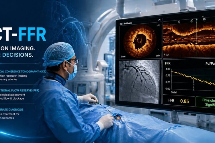
Optical coherence tomography (OCT) is a diagnostic procedure that is used during cardiac catheterization. It is a new, non-invasive imaging technique that generates volumetric angiography images in a matter of seconds.
OCT uses light and with OCT, doctors can obtain images of the blood vessels that are about the same as if they were looking under a microscope.
HOW DOES OCT WORK?
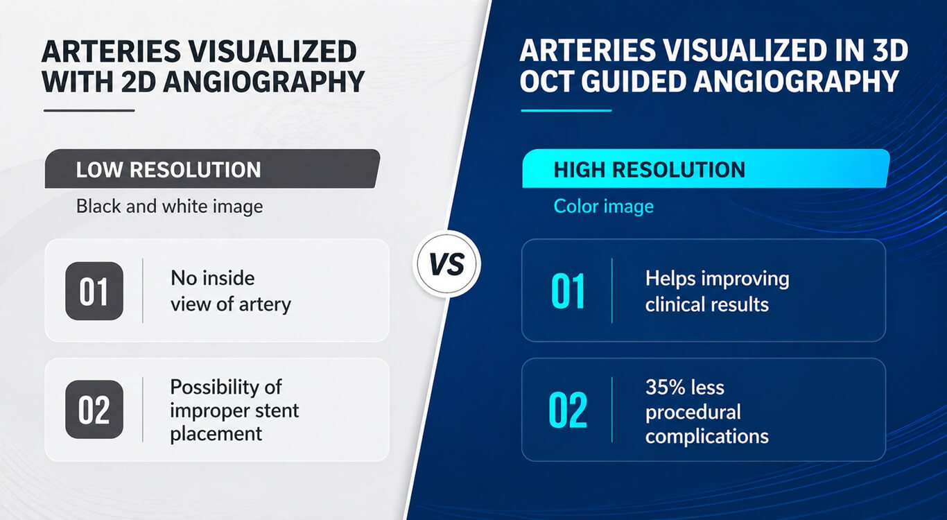
OCT uses near-infrared light to create images of the inside of the coronary arteries. The technique delivers very high-resolution images. In fact, OCT allows cardiologists to see the inside of an artery in 10 times more detail with 3D image than traditional angiography with 2D images.
BENEFITS WITH OCT
OCT also lets cardiologists clearly see the plaque inside an artery, find out how much fat or clot is inside an artery, and take precise measurements before and after placing stents.
FRACTIONAL FLOW RESERVE (FFR)
Fractional Flow Reserve (FFR) is a procedure to determine if a cardiac patient really needs a stent or bypass surgery or can be kept only on medicines avoiding any procedure. FFR technology not only saves lives while avoiding unnecessary surgery but also helps patients to save cost.
HOW DOES FFR WORK?
A very thin guide wire is inserted through a standard 4F or 5F diagnostic catheter during an angiogram. Because of the smaller size catheter necessary, this can be done as an outpatient procedure.
The special guide wire crosses the lesion and is able to measure the flow and pressure of the blood, after infusion of a hyperemic agent, such as adenosine. Results are displayed on a special monitor (left) along with the “FFR value”. [Source: PTCA.ORG]
Need more information about OCT & FFR, talk to us now.