Contact
Get in touch
Contact Form
Fill the form to contact & consult Dr. Tejas V. Patel.


Born in the historical land of Saurashtra, Dr Patel finished his school education from Junagadh. He completed his MBBS and MD (Medicine) from MP Shah Medical College, Jamnagar. He achieved 2nd rank in the Saurashtra University in MD.
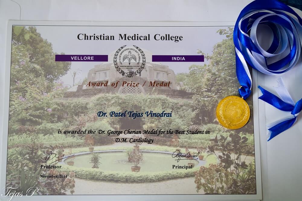
Dr Tejas V Patel pursued his super-speciality, DM in Cardiology from Christian Medical College (CMC), Vellore; which is considered as one of the best medical institutions in India. He was awarded the prestigious “Dr G M Cherian Gold Medal” for ‘Best Outgoing Student in DM Cardiology’. He worked as Assistant Professor in this premier institute, after he finished DM. He spent almost four years at CMC, where he gained extensive experience in clinical, interventional and research work in the field of Cardiology.
Dr. Tejas V Patel has wide experience of performing coronary angiographies and coronary angioplasties – majority performed through transradial approach. He is also trained for various peripheral vascular interventions including intervention in Takayasu arterities, Aortic aneurysm and dissection – EVAR and TEVAR procedures. Dr. Patel has independently performed various diagnostic and interventional cardiac procedures such as percutaneous ASD/VSD/PDA device closure, Balloon Mitral,Pulmonary,Aortic Valvulotomy and cardiac catheterization study with or without oxymetry of various congenital heart diseases.
He has done more than 20,000 transthoracic echocardiography including paediatric echocardiography, Tissue strain and strain rate echo, 3D echo and trans-oesophageal echo (TEE). As per the patients treated by him, he has been rated as one of the best cardiologist in Ahmedabad, Gujarat.
Dr. Tejas V. Patel has participated in several original research projects and many of his papers have been published at National and International levels in reputed journals including ‘Journal of American College of Cardiology (JACC)’. He has also given presentation at various national conferences. He was selected for zonal round (among top 10) in Torrent Young Scholar Award (TYSA) competition during both M.D. at Mumbai, 2009 and D.M. at Bangalore, 2013.
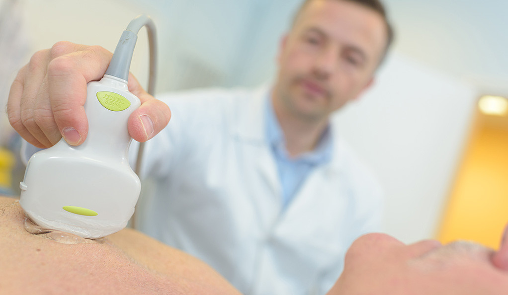
Echocardiography or simply an Echo is a sonography of the heart.
Echocardiography or simply an Echo is a sonography of the heart. It uses sound waves to create moving pictures of your heart. An echocardiogram can be done in the doctor’s office or a hospital. It requires undressing from the waist up and to lie down with turning left on an examination table or bed.
Your doctor may use this test to look at your heart’s structure and function in variety of diseases. This test may be needed if…
Depending on what information your doctor needs, you may have one of the following kinds of echocardiograms:
Transthoracic echocardiography (TTE): This is routinely done in which a probe (transducer) is placed on your chest. The probe sends special sound waves, called ultrasound, through your chest wall to your heart. As the ultrasound waves bounce off the structures of your heart, a computer in the echo machine converts them into pictures on a screen.
Trans-esophageal echocardiography (TEE): During this test, the transducer is attached to the tip of a flexible tube. The tube is introduced through your mouth into your food pipe (esophagus). TEE shows clearer pictures of your heart, because the probe is located closer to the heart. This allows your doctor to get more detailed pictures of your heart. Drug spray is given to your throat prior to the procedure for prevention of discomfort and pain.
Stress echocardiography: During this test, an echo is done both before and after your heart is stressed either by having you exercise or by injecting a medicine that makes your heart beat harder and faster. A stress echocardiogram is usually done to find out if you might have decreased blood flow to your heart due to possible blockage in the arteries supplying blood and oxygen to heart muscle.
Three-Dimensional (3D) echocardiography: A three-dimensional (3D) echo creates 3D images of your heart.
Coronary angiography is a test that uses special dye and special x-rays to show the insides of your coronary arteries.
Our heart pumps blood (and oxygen) to all the body organs. Heart muscle also needs oxygen for the function of pumping, which is supplied to the heart by Coronary arteries. Coronary angiography is a test that uses special dye and special x-rays to show the insides of your coronary arteries. The facility where such test is possible with special x-ray machine is called ‘Cath lab’ (catheterization laboratory).
In this procedure, a thin, hollow tube called a catheter is inserted into a blood vessel from your arm or groin. The tube is then advanced and threaded into your coronary arteries. Then dye is injected and x-ray pictures are taken while the dye is flowing through the coronary arteries. This helps doctors see blockages in the arteries. When the angiogram is over, the catheter is removed and pressure bandage is applied at the puncture site.
Prior to the angiography, you may need some blood tests and ECG (cardiogram). You may be asked to stop eating and drinking for few hours before the test. You may be asked to shave both groins and arms before the test. If you are pregnant, you need to inform the doctor prior to the test.
Before the test starts, you can be given a mild sedative, which helps you to be relaxed. You’re fully awake during the procedure, and it causes little or no pain. It takes only few seconds to finish this test. Generally the risk of serious complications is very low. You can be discharged on the same day of procedure. You should drink plenty of fluids to help flush the dye from your body.
Avoid strenuous activities and heavy lifting for several days. Your puncture site is likely to remain tender for a while. There can skin discoloration and a small swelling for few days at the puncture site.
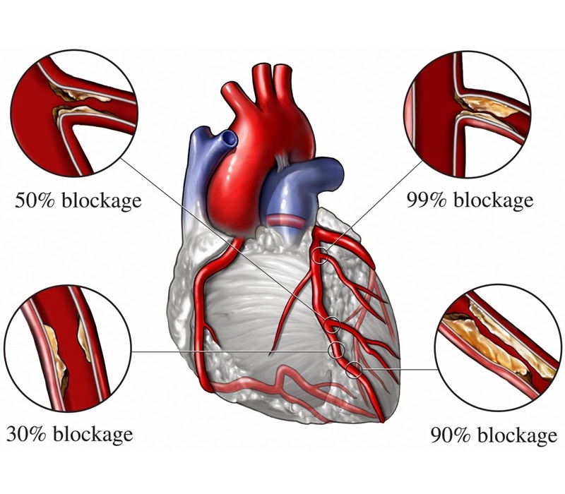
Coronary angiography is considered as a ‘gold-standard’ test to diagnose blocks in the artery of your heart. When cholesterol gets deposited in the wall of your coronary arteries (called as plaque), it causes narrowing of the lumen of arteries; which is called ‘Coronary artery disease (CAD)’. Narrowing of the artery causes reduction of oxygen-rich blood supply to your heart.
This can cause chest pain or discomfort called ‘Angina’. Sometimes a blood clot develops on the surface of plaque and it can partially or completely block blood flow through the coronary artery. This can compromise the viability of heart muscle and in few minute your heart muscle stops working. This can cause the most severe form of heart attack called ‘Myocardial infarction (STEMI)’. Coronary angiography is very useful test to diagnose above conditions.
Angioplasty is not a major surgery, it can be done from the same puncture (in arm or groin) done for angiography.
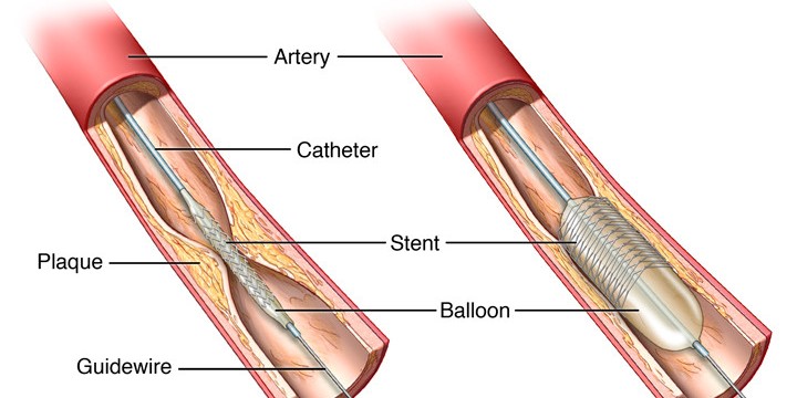
When cholesterol gets deposited in the wall of your coronary arteries (called as plaque), it causes narrowing of the lumen of arteries. This can reduce blood flow to your heart and cause chest pain (angina) or heart attack (myocardial infarction). Coronary angioplasty, also called PTCA (Percutaneous Transluminal Coronary Angioplasty) is the procedure which opens blocked arteries (by tiny balloon & stent) and restores normal blood flow to your heart muscle. In emergency situation like, heart attack (myocardial infarction/STEMI), angioplasty should be done as soon as possible and it is called as PAMI (Primary Angioplasty in Myocardial Infarction).
Angioplasty is not a major surgery, it can be done from the same puncture (in arm or groin) done for angiography. Patient remains fully awake throughout the procedure. Whole procedure is performed with the help of special x-ray machine in cardiac cath lab (cardiac catheterization laboratory). In this procedure, a thin, hollow tube called a catheter is inserted into a blood vessel from your arm or groin. The tube is threaded into your coronary arteries and tiny wire is advanced into the artery where the block is situated. The blocked artery is then opened by inflating a tiny balloon in it. Angioplasty is often combined with the placement of a stent. The whole procedure usually takes 30-60 minutes.
A stent is a short, metal mesh tube that acts like a scaffold to help keep your artery open. Stent remains in the body permanently. There are two main types of stent:
There are certain stents which are made from special materials that get dissolved or be absorbed in the body. This type of stent disappears completely from the body after few months.
You will usually remain hospitalized for one day while your heart is monitored and your medications are adjusted. You will normally be able to go home the day after the procedure. You will need to avoid heavy lifting, strenuous activities and driving for few days.
The most important thing is that you closely follow your doctor’s recommendations about your drugs, especially with blood-thinning medications such as Aspirin and clopidogrel or similar medications like Ticagrelor/Prasugrel. Those who have had stent placement will need a blood-thinning medication for a year or longer in some cases. If you have any doubts or if you need any surgery, talk to your cardiologist before stopping any of these medications.
A pacemaker is a small device, smaller than the size of your fist; that’s placed under the skin near your heart to help control your heartbeat.
Every heart has its own internal electrical system and natural pacemaker that regulates the rate and rhythm of your heartbeats. With each heartbeat, an electrical signal spreads from the top of your heart to the bottom. As the signal travels, it causes the heart to contract and pump blood.
However, sometimes your heart beats too slowly that it may not be able to pump enough blood to the body. A pacemaker can correct this problem. It is a small device that sends electrical impulses to the heart muscle to maintain adequate heart rate and rhythm. A pacemaker is a small device, smaller than the size of your fist; that’s placed under the skin near your heart to help control your heartbeat.
Doctors may recommend pacemakers to you for many reasons. The most common reasons are very low heart rate and heart block. Heart block is a problem in the electrical system of the heart so that electrical signals cannot reach from upper chambers to the lower chambers of your heart. As a consequence pumping of your heart is reduced. Heart block can happen as a consequence of advanced aging, severe heart attack, or certain neuromuscular disorders.
Pacemakers can be temporary or permanent. Temporary pacemakers are used to treat short-term heart problems and during emergencies. They might be used until your doctor can implant a permanent pacemaker or until a temporary condition goes away.
There are different types and modes of pacemaker. Your doctor will decide what type of pacemaker is best for you based on your heart condition.
It is a special type of pacemaker, which is useful to control very fast heart rate which can occur in certain diseases.
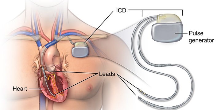
Implantable Cardioverter Defibrillator (ICD): It is a special type of pacemaker, which is useful to control very fast heart rate which can occur in certain diseases. Very fast heart rate of lower chamber of the heart (ventricular tachycardia/fibrillation) is very serious and life threatening condition. If it is not treated promptly, patient can become unconscious or even die in few minutes. ICD is a special device similar to pacemaker can immediately deliver electrical impulse or shock to the heart when it detects this serious rhythm problem.
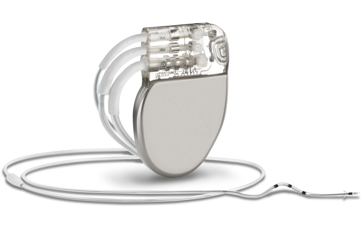
Biventricular pacemaker / Cardiac Resynchronization Therapy (CRT): It contains three leads – two in upper and lower chambers of right side of the heart and third placed in left side of the heart. It is useful in the patient of heart failure. A biventricular pacemaker paces both ventricles so that all or most of the ventricular muscle pumps together. This allows the heart to pump blood more effectively.
Physical Activity: Do not hold your arm above shoulder level for 3-4 weeks after pacemaker implantation. Avoid activities that require pushing or pulling heavy objects. Ask your doctor how much and what kinds of physical activities are safe for you. Self-driving should be avoided if you have implanted ICD.
MRI: Avoid standing near strong magnetic field including MRI rooms if you have pacemaker implanted. All pacemakers are not MRI compatible and you cannot undergo MRI if you have pacemaker which is not compatible with MRI. Even though you have MRI compatible pacemaker, consult your cardiologist before undergoing any MRI.
Electrical Interference:
Once your pacemaker is implanted, the battery should last 5-10 years, depending upon how frequently you need pacing. When a pacemaker’s battery wears out, your doctor will replace the generator along with the battery. Your doctor can tell you whether your pacemaker or its wires need to be replaced after interrogation of your pacemaker.
Balloon Valvuloplasty (or valvulotomy) means dilation of narrowed cardiac valve with balloon.
Balloon Valvuloplasty (or valvulotomy) means dilation of narrowed cardiac valve with balloon. This modality of treatment useful in certain diseases of valves of the heart in which valve stenosis occurs either due to birth defect or due to acquired inflammation. This procedure is usually done by a small injection from your groin region under local anesthesia. Mild sedation is given to you for pain relief. You remain conscious during the procedure; unlike valve replacement surgery which is an open heart surgery and requires general anesthesia.
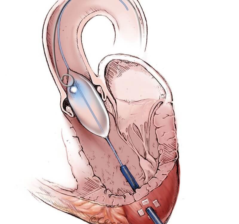
In this procedure a long, thin tube (catheter) is inserted from your groin. Then a balloon is advanced to your heart and passed across the diseased narrowed valve. Now the balloon is inflated to open or stretch the valve. The balloon is then deflated and it is removed along with the catheter from your body. Balloon valvuloplasty relieves many symptoms of heart valve disease, but may not completely cure it.
Balloon valvuloplasty is useful for all 4 cardiac valves named Mitral, Aortic, Pulmonary and Tricuspid. It is commonly done in rheumatic heart disease involving mitral valve causing mitral stenosis (narrowing of mitral valve). Balloon aortic valvuloplasty and ballon pulmonary valvuloplasty are commonly performed in children.
If you have any heart valve problem, you should consult your cardiologist. Cardiologist will perform Echocardiography to look at the problem in the valve and then can advise whether you should get benefit of balloon valvuloplasty and whether it is an appropriate treatment for your condition.
Peripheral angiography is used to check condition of blood vessels supplying your legs and arms.
A peripheral angiography is a test similar to coronary angiography. Unlike coronary angiography which is used to look the condition of artery supplying your heart; peripheral angiography is used to check condition of blood vessels supplying your legs and arms. This test uses special x-ray machine and it is performed in cath lab.
Peripheral angiogram is useful test for the diagnosis of peripheral artery disease (PAD). PAD is a disease in which cholesterol and/or blood clots cause narrowing of the lumen of the artery supplying blood to your the legs or arms. Such blocks in the artery cause muscle pain or cramping in legs or arms on activity, such as walking or climbing stairs, and disappears after a few minutes of rest (intermittent claudication).
Once the disease progresses, you may feel pain even at rest. Critical narrowing of the artery can cause ulcer or gangrene formation in your leg.
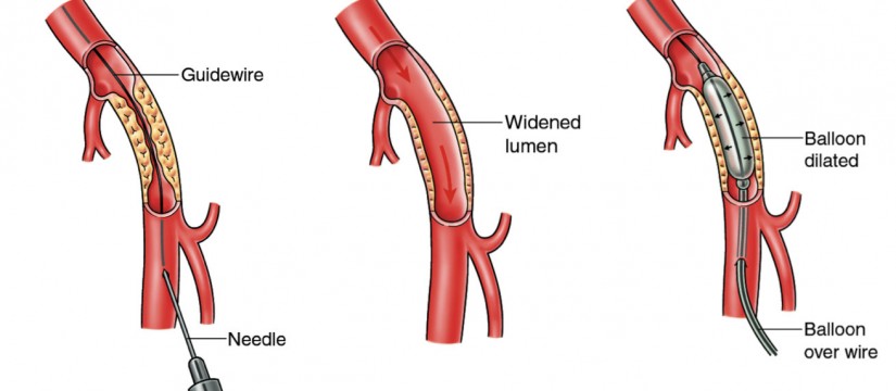
Treatment for peripheral artery disease includes special drugs that reduce the pain in your leg while walking or climbing stairs. If symptoms are not relieved with the medications then angioplasty or surgery is indicated. In peripheral angioplasty, a catheter (thin hollow tube) is inserted through your groin. Then in this catheter a balloon is advanced in the blocked artery.
The balloon is then inflated; this widens the artery and reduces the block. A stent (a small mesh tube) may be placed in the artery during angioplasty. A stent helps keep the artery open and prevent re-occlusion.
Sometimes your artery may be blocked due to blood clot in it. In such situation injection of a clot-dissolving drug (clot busters) into your artery through a catheter (thin hollow tube) is done to break the clot and to restore the blood flow in the blocked artery.
Recent Comments