Peripheral-Angiography-Angioplasty-min-825x360

PERIPHERAL ANGIOGRAPHY AND ANGIOPLASTY

Peripheral angiography is used to check condition of blood vessels supplying your legs and arms.

PERIPHERAL ANGIOGRAPHY AND ANGIOPLASTY

A peripheral angiography is a test similar to coronary angiography. Unlike coronary angiography which is used to look the condition of artery supplying your heart; peripheral angiography is used to check condition of blood vessels supplying your legs and arms. This test uses special x-ray machine and it is performed in cath lab.

WHEN TO DO PERIPHERAL ANGIOGRAPHY?


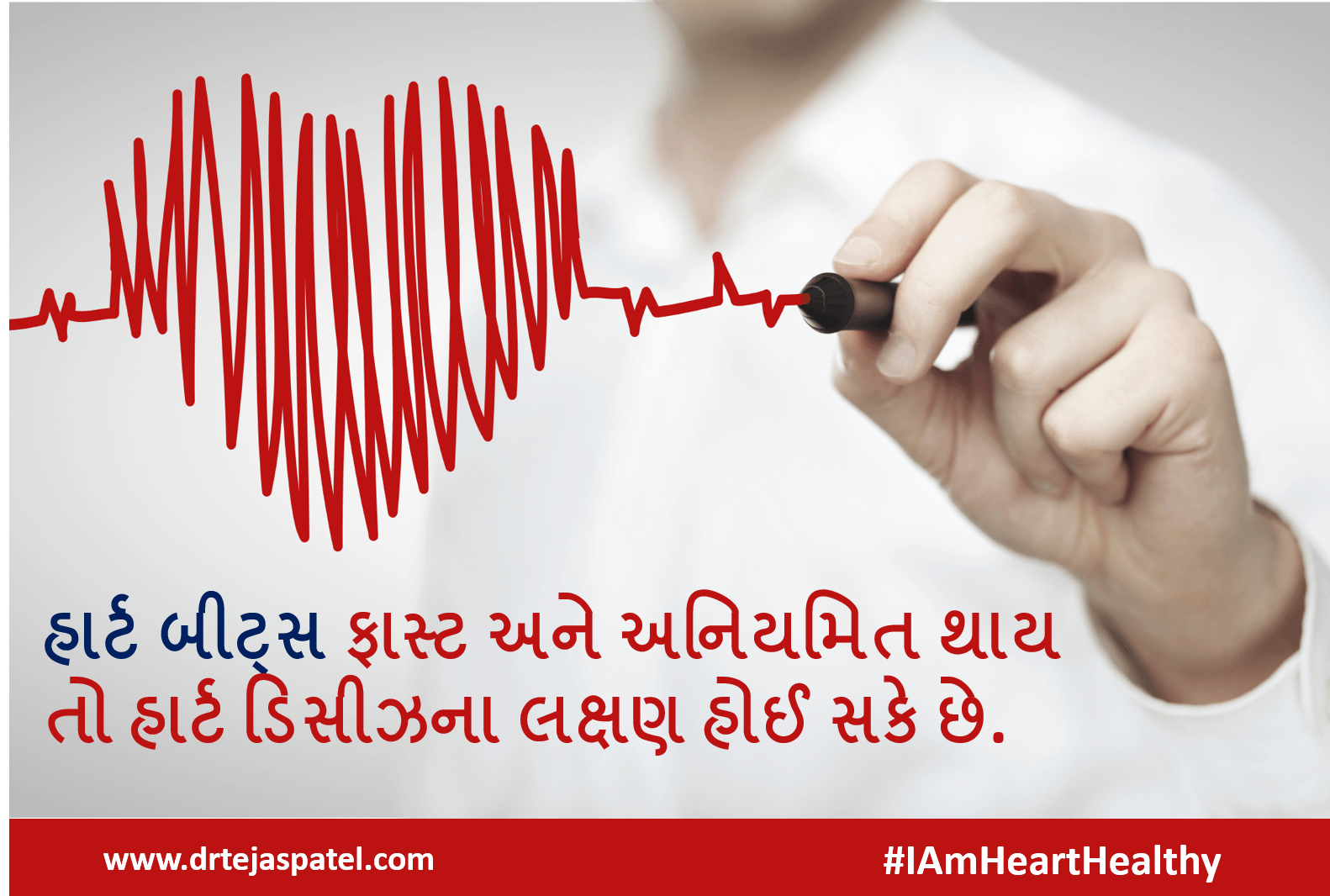
Peripheral angiogram is useful test for the diagnosis of peripheral artery disease (PAD). PAD is a disease in which cholesterol and/or blood clots cause narrowing of the lumen of the artery supplying blood to your the legs or arms. Such blocks in the artery cause muscle pain or cramping in legs or arms on activity, such as walking or climbing stairs, and disappears after a few minutes of rest (intermittent claudication).

Once the disease progresses, you may feel pain even at rest. Critical narrowing of the artery can cause ulcer or gangrene formation in your leg.

PERIPHERAL ANGIOPLASTY


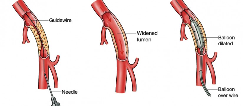
Treatment for peripheral artery disease includes special drugs that reduce the pain in your leg while walking or climbing stairs. If symptoms are not relieved with the medications then angioplasty or surgery is indicated. In peripheral angioplasty, a catheter (thin hollow tube) is inserted through your groin. Then in this catheter a balloon is advanced in the blocked artery.

The balloon is then inflated; this widens the artery and reduces the block. A stent (a small mesh tube) may be placed in the artery during angioplasty. A stent helps keep the artery open and prevent re-occlusion.

CATHETER-DIRECTED THROMBOLYSIS (CDT)


Sometimes your artery may be blocked due to blood clot in it. In such situation injection of a clot-dissolving drug (clot busters) into your artery through a catheter (thin hollow tube) is done to break the clot and to restore the blood flow in the blocked artery.

RENAL/CAROTID ANGIOPLASTY

Renal angioplasty restores blood flow through the renal artery and improves blood flow to your kidney.

RENAL & CAROTID ANGIOPLASTY

RENAL ANGIOPLASTY

Renal artery stenosis is the narrowing of the arteries that supply blood to the kidneys. It can lead to uncontrolled hypertension, which should be treated initially with medications. If you have renal artery stenosis and blood pressure remains uncontrolled or complications such as pulmonary edema or worsening kidney function develop, angioplasty with stenting should be performed. Renal angioplasty restores blood flow through the renal artery and improves blood flow to your kidney. Renal angioplasty can be done by a small puncture done from your groin or sometimes from left arm. Initially the blocked artery is opened by inflating a tiny balloon in it; it is followed by permanent placement of a stent. This procedure is very similar to coronary angioplasty.

CAROTID ANGIOPLASTY

Cholesterol deposition (plaque) can narrow or block your carotid arteries which supply blood to the brain. When one or both of your carotid arteries are narrowed, it can make it hard for blood to flow to the brain. Sometimes blood clot develops in that narrowed part and may dislodge to the brain. This blood clot in the brain can block the artery in your brain and that results in stroke and paralysis. Carotid artery angioplasty and stenting may improve blood flow to your brain and lower the risk of having a stroke.

Your doctor may suggest for carotid artery stenting if:

  • You have had a mild stroke or transient ischemic attacks (TIAs) in the past 6 months and your carotid artery is narrowed by 70% or more.
  • You have a low risk for complications from the procedure.
  • You have a high risk of complications from carotid artery surgery.

Carotid angioplasty is performed by a small puncture in your groin. The procedure is similar to coronary angioplasty. Additionally, an umbrella-shaped filter (embolic protection device) is placed beyond the block during the procedure; it helps to catch any debris that may break off from the narrowed area of artery during ballooning and stenting.

TAVR1

TAVR – Transcatheter Aortic Valve Replacement

Lorem ipsum dolor sit amet, conse tetur adipisicing elit, sed do.

TAVR – Transcatheter Aortic Valve Replacement

WHAT IS TAVR? (Also called TAVI)


Transcatheter aortic valve replacement (TAVR) is a minimally invasive procedure to replace a narrowed aortic valve that fails to open properly (aortic valve stenosis). Transcatheter aortic valve replacement is sometimes called transcatheter aortic valve implantation (TAVI).

TAVR may be an option for people who are considered at intermediate or high risk of complications from surgical aortic valve replacement. TAVR may also be indicated in certain people who can’t undergo open-heart surgery.

WHO BENEFITS MOST FROM TAVR?


TAVR may be an option if you have aortic stenosis that causes signs and symptoms. For instance, people who are candidates for TAVR may include those who are considered at intermediate or high risk of complications from surgical aortic valve replacement. Conditions that may increase the risk of surgical aortic valve replacement include lung disease or kidney disease — which increase your risk of complications during surgical aortic valve replacement.

HOW IS TAVR OR TAVI DIFFERENT FROM THE STANDARD VALVE REPLACEMENT?


Usually valve replacement requires an open heart procedure with a “sternotomy”, in which the chest is surgically separated (open) for the procedure. The TAVR or TAVI procedures can be done through very small openings that leave all the chest bones in place. The TAVR procedure is performed using one of two different approaches, allowing the cardiologist or surgeon to choose which one provides the best and safest way to access the valve:

Entering through the femoral artery (large artery in the groin), called the transfemoral approach, which does not require a surgical incision in the chest, or
Using a minimally invasive surgical approach with a small incision in the chest and entering through a large artery in the chest or through the tip of the left ventricle (the apex), which is known as the transapical approach.

TAVR/TAVI – Latest Treatment of Aortic Valve/Heart Valve


EVAR AND TEVAR FOR AORTIC DISSECTION/ANEURYSM

are newer techniques used to treat pathology of the aorta, like aortic aneurysm and dissection.

EVAR & TEVAR FOR AORTIC DISSECTION & ANEURYSM

AORTIC DISSECTION

Aorta is the largest artery in the body and arising directly from the heart. Aortic dissection occurs when a tear develops in the wall of the aorta. As the tear extends along the wall of the aorta, blood can flow in between the layers of the blood vessel wall (dissection). This can lead to aortic rupture or decreased blood flow to organs. In most cases, the symptoms begin suddenly, and include severe chest pain. The pain may mimics heart attack. The pain is usually described as tearing or stabbing and radiating to the shoulder blades.

There are 2 types of aortic dissection:

  • Type A – aortic dissection which involves ascending aorta and arch
  • Type B – aortic dissection which involves descending aorta

Aortic dissection is a medical emergency requiring immediate treatment. Treatment may include surgery or endovascular repair depending on the location of the dissection.

AORTIC ANEURYSM

Aortic aneurysm is a bulge or ballooning in its wall. If an aneurysm grows large, it can burst and cause dangerous bleeding or even death.

There are 2 types of aortic aneurysm:

  • Thoracic aortic aneurysms – these involves the part of the aorta running through the chest cavity
  • Abdominal aortic aneurysms – these involves the part of the aorta passing through the abdomen

CT aortogram is helpful in the diagnosis of this condition. Small aortic aneurysms which are not causing any symptoms are monitored over time until they become large. When an aortic aneurysm is large or associated with symptoms, it should be repaired by surgery or by endovascular approach like EVAR or TEVAR.

EVAR & TEVAR

EVAR (abdominar EndoVascular Aneurysm/Aortic Repair) and TEVAR (Thoracic EndoVascular Aneurysm/Aortic Repair) are newer techniques used to treat pathology of the aorta, like aortic aneurysm and dissection. The procedure involves the placement of an expandable stent graft within the aorta to treat aortic disease without operating directly on the aorta. Incision is made in both side of groin to cannulate femoral arteries.

It is similar to the approach used for coronary angiography and angioplasty. Then stent-graft is introduced through the femoral artery in the groin and with the use of x-ray guidance and specially-designed instruments, the aneurysm can be repaired from inside the aorta by deploying the stent-graft.

The stent graft provides a new channel for the blood to flow through, reducing the pressure on the diseased area of the artery and helping to prevent a rupture. The recovery time for endovascular repair is less than the recovery time for open abdominal or open chest repair.

Surviving A Stemi Heart Attack – Dr Tejas V. Patel CIMS

An ST-segment Elevation Myocardial Infarction (STEMI) is the most serious form of heart attack in which a coronary artery is completely blocked, resulting a large part of the heart muscle being unable to receive blood. Let’s see how to survive this most dangerous form of heart attack.

Categories:

7 સંકેતો જે હાર્ટની તકલીફ હોવા તરફ ઈશારો કરે છે

Info 2
INfo 2
Info 7
Info 6
Info 5
Info 4
Info 3

Categories:

10 Heart Healthy Habits

Habits influence how we live our lives and our health. So what are the good habits you follow in your daily life for your heart? Here’re some of the good and heart healthy habits that everyone needs to focus for a healthy heart.
Heart_Healthy_HabitsFollow our Facebook Page (/TejasPatel.Cardiologist) & learn more ways and health tips to prevent heart disease at any age 20s, 30s, 40s, 50s and beyond.

Categories:

Whipped Cream Cheese: Is It Good For Heart?

Who don’t like cheese? However it has more fat content, which makes people skeptical about eating cheese. Cream cheese comes in numerous forms: brick, regular, whipped, light, fat-free and Neufchatel and a dozen more or the type.

And among different kinds of cheeses, is cream cheese healthy?

It totally depends on which kind you choose. Whipped cream cheese could be a better option when it comes to a healthy heart. So if you are a cream cheese lover, give it a touch of whip.
Regular cream cheese has a fair amount of fat, especially the artery-clogging kind, for a pretty moderate serving. But cream cheese can still grace your morning whole-wheat bagel. Reach for the whipped type in the dairy case. You can save about half the calories and saturated fat.
Whipped cream cheese incorporates air (from whipping) so it seems as if you’re eating more. Two tablespoons have 80 calories, 8 grams of fat and 5 grams of saturated fat. (source: blog.foodnetwork.com)
The verdict: If you are a cream cheese lover, give it a touch of whip.To make a little go even further, have some whipped cream cheese bagels. Beyond the traditional uses, cream cheese can enhance the taste of many healthy recipes like mashed potatoes, fruit pizza, etc. As always, the key is to check for your heart and say #IAmHeartHealthy.

Categories: